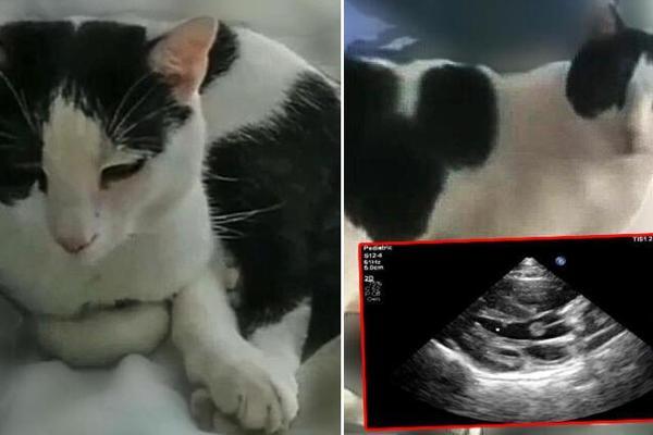
Heart valve tumor detected in cat, first time in world
AFYONKARAHİSAR


A cat taken to the animal hospital in the central province of Afyonkarahisar with the complaint of respiratory distress has been diagnosed with a heart valve tumor, the first in the world.
The tabby cat was taken to the animal hospital of Afyon Kocatepe University (AKÜ) by its owner.
Following the examination of the radiological images, the veterinarians who examined the cat found papillary fibroelastoma, a heart valve tumor, identified by echocardiography. The cat, whose condition is currently stable, has been put on medication.
Examining the images, Turan Civelek, the dean of the veterinary medicine faculty, determined that the tumor was rare and was diagnosed in a cat for the first time.
Noting that heart diseases are frequently encountered in cats and dogs of all ages, he emphasized that cardiac tumors are rare in cats.
The tumor in the tabby was benign, the professor said. “However, these types of tumors carry the risk of thromboembolism. It can cause complications such as stroke or infarction. Symptoms are often unclear and may vary depending on the size or structure of the mass.”
Civelek said they made a report and published it in Kocatepe Veterinary Journal, the university’s organ, in a bid to produce a reference work for their colleagues.
“As a result of our research, we have seen that there is no previously reported case of fibroelastoma in cats in the scientific world. It is also gratifying for us that this is a first. This report is important in terms of guiding veterinarians and describing the case in detail.”
Pointing out that early diagnosis saves lives, Civelek said, “Animal lovers living with ‘friends with paws’ should always be careful in terms of possible heart diseases. Routine heart examinations at early ages and again after middle age are important in terms of maintaining their life quality and early diagnosis.”
The previously undiagnosed diseases can be detected with modern diagnostic equipment, Civelek said, underlining that the use of blood analysis, x-ray, endoscopy and ultrasonography for veterinary medicine are now among the routine applications.
“Computed tomography and even MRI are now used in diagnosis in many centers in our country,” the expert added.
Although rare in veterinary medicine, cardiac tumors can cause life-threatening complications. However, as presenting clinical signs in patients with cardiac tumors are usually nonspecific, a low level of suspicion often hampers diagnosis.
Reports from owners may include hyporexia, lethargy, weakness, neurologic signs, vomiting, and abdominal distention.
